


Home / Training / Manuals / Atlas of breast cancer early detection / Cases
Atlas of breast cancer early detection
Go back to the list of case studies
.png) Click on the pictures to magnify and display the legends
Click on the pictures to magnify and display the legends
| Case number: | 095 |
| Age: | 58 |
| Clinical presentation: | Postmenopausal woman with average risk of developing breast cancer presented with a lump in the left breast. Examination revealed a 1.5 cm lump in the retroareolar region of the left breast. |
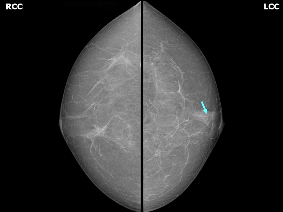
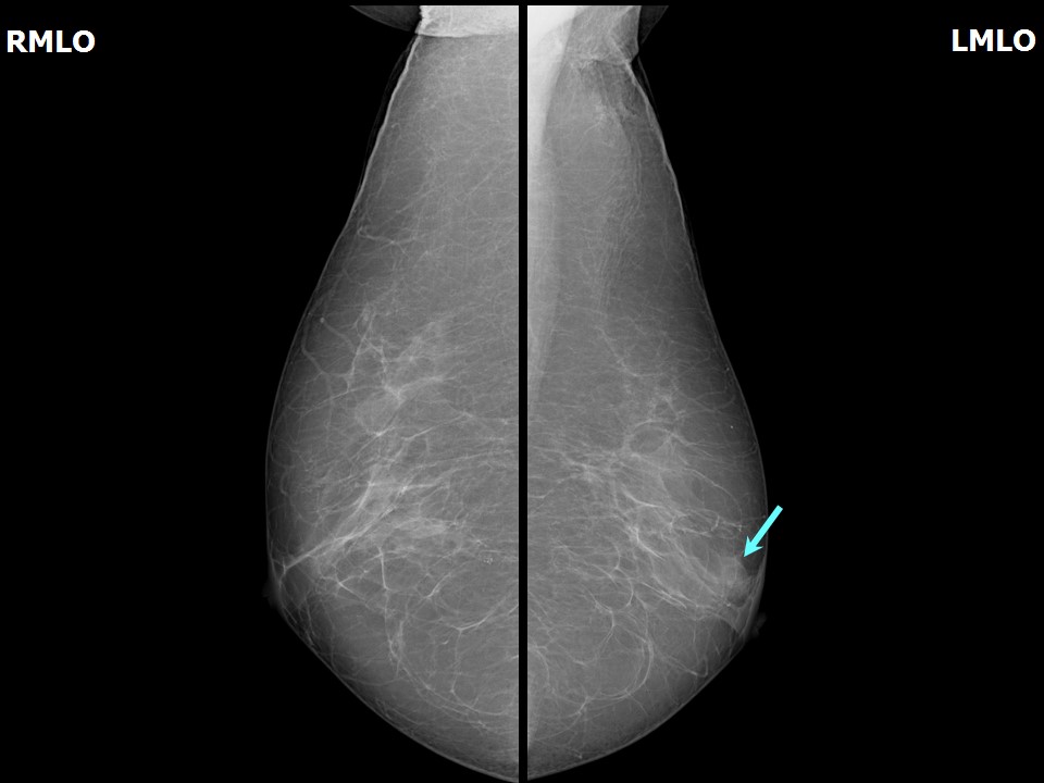
Mammography:
 |  |
 | 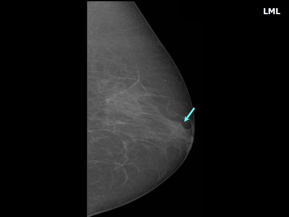 |
| Breast composition: | ACR category a (the breasts are almost entirely fatty) | Mammography features: |
| ‣ Location of the lesion: | Left breast, central portion of the breast, central zone, anterior third |
| ‣ Mass: | |
| • Number: | 1 |
| • Size: | 1.2 × 0.9 cm |
| • Shape: | Irregular |
| • Margins: | Indistinct |
| • Density: | Equal |
| ‣ Calcifications: | |
| • Typically benign: | None |
| • Suspicious: | None |
| • Distribution: | None |
| ‣ Architectural distortion: | None |
| ‣ Asymmetry: | None |
| ‣ Intramammary node: | None |
| ‣ Skin lesion: | None |
| ‣ Solitary dilated duct: | None |
| ‣ Associated features: | None |
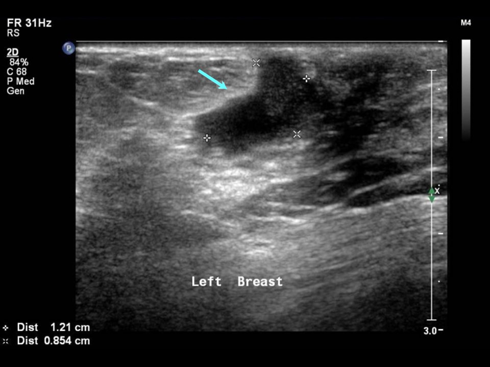
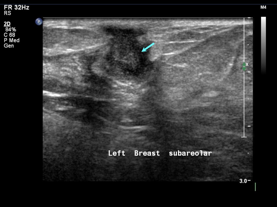
Ultrasound:
 |  |
 |
| Ultrasound features: Left breast, central portion of the breast | |
| ‣ Mass | |
| • Location: | Left breast, central portion of the breast |
| • Number: | 1 |
| • Size: | 1.2 × 0.8 cm |
| • Shape: | Irregular |
| • Orientation: | Not parallel |
| • Margins: | Indistinct |
| • Echo pattern: | Heteroechoic |
| • Posterior features: | No posterior features |
| ‣ Calcifications: | None |
| ‣ Associated features: | Vessels in rim |
| ‣ Special cases: | None |
BI-RADS:
BI-RADS Category: 4A (low level of suspicion for malignancy)Further assessment:
Further assessment advised: Referral for cytologyCytology:
 |
| Cytology features: | |
| ‣ Type of sample: | FNAC (solid lesion) |
| ‣ Site of biopsy: | |
| • Laterality: | Left |
| • Quadrant: | Retroareolar lump |
| • Localization technique: | Palpation |
| • Nature of aspirate: | 0.2 mL of thick whitish material |
| ‣ Cytological description: | Smear shows many neutrophils and foamy macrophages. Epithelioid histiocytes are seen in large conglomerates |
| ‣ Reporting category: | Benign |
| ‣ Diagnosis: | Granulomatous inflammation |
| ‣ Comments: | None |
Histopathology:
Breast lumpectomy
 |  |
| Histopathology features: | |
| ‣ Specimen type: | Breast lumpectomy |
| ‣ Laterality: | Left |
| ‣ Macroscopy: | Cut surfaces of the serial sections do not show any tumour nodules. Soft whitish areas are noted in the adipose tissue |
| ‣ Histological type: | Section shows granulomatous mastitis with epithelioid cell conglomerates, Langhans giant cells, and peripheral cuff of lymphocytes. Some dilated ducts with inspissated secretions and surrounded by chronic inflammation are seen |
| ‣ Histological grade: | |
| ‣ Mitosis: | |
| ‣ Maximum invasive tumour size: | |
| ‣ Lymph node status: | |
| ‣ Peritumoural lymphovascular invasion: | |
| ‣ DCIS/EIC: | |
| ‣ Margins: | |
| ‣ Pathological stage: | |
| ‣ Biomarkers: | |
| ‣ Comments: | Negative for acid-fast bacilli |
Case summary:
| Postmenopausal woman presented with left breast lump. Diagnosed as left breast subareolar tubular irregular lesion, BI-RADS 4A on imaging and as granulomatous mastitis on cytology and histopathology. |
Learning points:
|